42 ribosome diagram with labels
› proteins › proteinProtein Targeting (With Diagram) | Molecular Biology ADVERTISEMENTS: Let us make an in-depth study of the protein targeting. After reading this article you will learn about: 1. Introduction to Protein Targeting 2. Signal Sequence 3. Transport of Proteins into ER 4. Signal Sequence Recognition Mechanism 5. Role of Golgi Complex in Protein Transportation 6. Transport of Proteins from Golgi to Lysosomes 7. […] Ribosome and protein synthesis, diagram - Science Photo Library Diagram showing protein synthesis in cells (translation). Messenger ribonucleic acid (mRNA, blue with coloured nucleotides) is read by a ribosome (pink). The molecules of transfer RNA (tRNA, key-shaped) each bring an amino acid (orange dot) to bind to the ribosome's protein synthesis site.
Structure of Ribosome - Biology Wise Diameter of Ribosome is 20nm. Their composition can be divided into two parts - 2/3 part of r-RNA (ribosomal RNA) and 1/3 part RNP (Ribosomal protein or Ribonuclep protein). Polypeptide chain is fabricated by translating mRNA (messenger RNA) with the aid amino acids that tRNA (transfer RNA) delivers.

Ribosome diagram with labels
Protein Targeting (With Diagram) | Molecular Biology ADVERTISEMENTS: Let us make an in-depth study of the protein targeting. After reading this article you will learn about: 1. Introduction to Protein Targeting 2. Signal Sequence 3. Transport of Proteins into ER 4. Signal Sequence Recognition Mechanism 5. Role of Golgi Complex in Protein Transportation 6. Transport of Proteins from Golgi to Lysosomes 7. […] Ribosomes: Definition, Types, Structure, Functions Figure: Diagram of a 70S ribosome . The molecular weight of the 70S ribosome is 2.5×10 6 daltons and the dimension of each ribosome is about 200-290 Å × 170-210 Å. 80S Ribosome. They are relatively larger in size than 70S ribosomes and are mainly found in the cytoplasm of eukaryotic cells (plant and animal cells). ... Mastering Biology Chapter 14 Flashcards - Quizlet Place the blue labels in their proper locations on this diagram showing the process of transcription. Then, use the pink labels to identify the corresponding RNA nucleotide that belongs in each pink target. Pink labels can be used once, more than once, or not at all. Drag the appropriate labels to their respective targets.
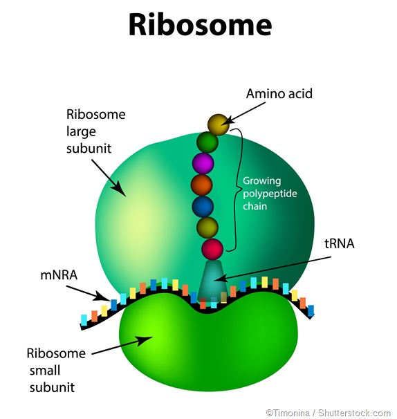
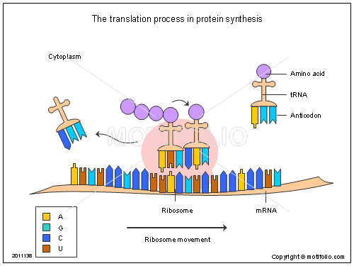
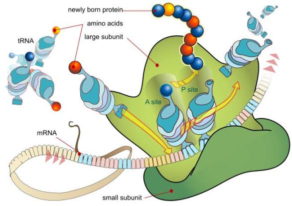
Ribosome diagram with labels. Active Ribosome Profiling with RiboLace - PubMed Ribosome profiling, or Ribo-seq, is based on large-scale sequencing of RNA fragments protected from nuclease digestion by ribosomes. Thanks to its unique ability to provide positional information about ribosomes flowing along transcripts, this method can be used to shed light on mechanistic aspects … Active Ribosome Profiling with RiboLace Bio 1113 - Unit 11 - Gene Expression Flashcards | Quizlet In the following diagram of a ribosome, assign the correct labels: Label 1: a tRNA attached to a polypeptide is found in this area of the ribosome Label 2: a tRNA attached to a single amino acid enters here Label 3: a tRNA that is not attached to anything exits here Label 4: a tRNA molecule Label 5: growing polypeptide Label 6: mRNA being ... Plant Cell Diagram | Science Trends A plant cell diagram, like the one above, shows each part of the plant cell including the chloroplast, cell wall, plasma membrane, nucleus, mitochondria, ribosomes, etc.A plant cell diagram is a great way to learn the different components of the cell for your upcoming exam. Plants are able to do something animals can't: photosynthesize.Plant cells are able to do this because plant cells have ... Protein Synthesis Labeling.pdf - 1. Label the diagram.... Protein DNA Amino Acid mRNA Codon tRNA Ribosome Anticodon 2. Label the diagram. Protein Amino Acid Large subunit - rRNA tRNA mRNA codon small subunit rRNA 3. What is the role of mRNA in the process of protein synthesis? The role of mRNA in protein synthesis is that it carries copies of genetic instructions (that tell the cell how to assemble ...
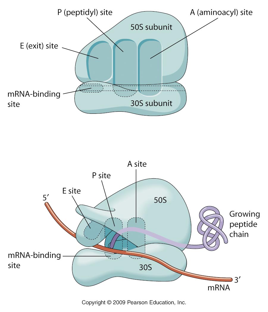
Solved The ribosome in the diagram is in the process of | Chegg.com The ribosome in the diagram is in the process of synthesizing a protein using directions transcribed from the DNA. Use the labels to identify each of the structures involved in translation and protein synthesis. Question: The ribosome in the diagram is in the process of synthesizing a protein using directions transcribed from the DNA. Ribosome - Wikipedia Prokaryotic ribosomes are around 20 nm (200 Å) in diameter and are composed of 65% rRNA and 35% ribosomal proteins. Eukaryotic ribosomes are between 25 and 30 nm (250-300 Å) in diameter with an rRNA-to-protein ratio that is close to 1. quizlet.com › 187533799 › campbell-ap-biologyCampbell Ap Biology Mastering Biology Chapter 17 Course Work Drag the white and purple labels to the white targets to indicate what each mutant mRNA codon codes for. (You will probably need to consult the codon table for mRNA .) Drag the pink labels to the pink targets to indicate the type of mutation. Drag the blue labels to the blue targets to indicate the effect on the polypeptide's primary structure. Cell Organelles- Definition, Structure, Functions, Diagram In the case of prokaryotic cells, the ribosomes are of the 70S with the larger subunit of 50S and the smaller one of 30S. Eukaryotic cells have 80S ribosomes with 60S larger subunit and 40S smaller subunit. Ribosomes are short-lived as after the protein synthesis, the subunits split up and can be either reused or remain broken up.
Ribosomes: Definition, Structure, Types, Functions and Diagram Ribosomes: Definition, Structure, Types, Functions and Diagram. The name ribosome is derived from two words: ribonucleic acid and 'somes', which is derived from the Greek word soma, which meaning "body.". Ribosomes are dense spheroidal particles with a diameter of 150 to 200 A0 found in most bacterial and eukaryotic cells. Cambridge Assessment International Education Cambridge 1 The diagram shows a leaf on a plant. Sun water from the soil carbon dioxide from the air simple sugars made in the leaf Which characteristic of life is represented by this diagram? A excretion B nutrition C respiration D sensitivity 2 The diagram shows how Homo sapiens (modern people) could have evolved from earlier ancestors. Homo habilis ... Prokaryotic Cells - BioNinja Prokaryotes are organisms whose cells lack a nucleus ('pro' = before ; 'karyon' = nucleus). They belong to the kingdom Monera and have been further classified into two distinct domains: Archaebacteria – found in extreme environments like high temperatures, salt concentrations or pH (i.e. extremophiles); Eubacteria – traditional bacteria including most known pathogenic forms … Biology: parts of a ribosome Diagram | Quizlet Start studying Biology: parts of a ribosome. Learn vocabulary, terms, and more with flashcards, games, and other study tools.
Cell Biology | Biology Images and Diagrams | Biology.Jpeg
What Are Ribosomes? - Definition, Structure and its Functions Ribosomes are located inside the cytosol found in the plant cell and animal cell. The ribosome structure includes the following: It is located in two areas of cytoplasm. Scattered in the cytoplasm. Prokaryotes have 70S ribosomes while eukaryotes have 80S ribosomes. Around 62% of ribosomes are comprised of RNA, while the rest is proteins.

Animal Cell diagram label
Ribosomes: Structure, Composition, and Assembly (With Diagram) Ribosomes in the cytoplasm of eukaryotic cells have a sedimentation coefficient of about 80 S (MW about 4.5 x 10 6) and are composed of 40 S and 60 S subunits. In prokaryotic cells, ribosomes are typically about 70 S (MW about 2.7 x 10 6) and are formed from 30 S and 50 S subunits.

Microbiology101506
Labeled Plant Cell With Diagrams | Science Trends The ribosomes are created in the nucleolus of the cell. Ribosomes are made out of two smaller subunits, a large ribosomes subunit and a small ribosomal subunits. The transfer RNA or tRNA encodes the correct series of genetic instructions into the mRNA or messenger RNA, which is what ensures that the right proteins are created.

Ribosome: Types, Structure and Functions | Biology Edu Care
Structure of Ribosome (With Diagram) - Biology Discussion A bacterial ribosome is about 250 nm in diameter and consists of two subunits, one large and one small. Both subunits consist of one or more molecules of rRNA and an array of ribosomal proteins. ADVERTISEMENTS: Association of two subunits is called mono-some. The structure of prokaryotic ribosome is given in the figure 8.2 B.
Functions and Parts of the Digestive and Urinary Systems
cdn.savemyexams.co.uk › uploads › 2022/03/0970_s19Cambridge Assessment International Education Cambridge ... 1 The diagram shows a leaf on a plant. Sun water from the soil carbon dioxide from the air simple sugars made in the leaf Which characteristic of life is represented by this diagram? A excretion B nutrition C respiration D sensitivity 2 The diagram shows how Homo sapiens (modern people) could have evolved from earlier ancestors. Homo habilis ...
Chapter 2: cells Flashcards | Easy Notecards
Oxford Cambridge and RSA Friday 16 October 2020 – Morning ribosome Fig. 1.2. 3 ... State three changes, other than to the labels, to Fig. 1.2 that the student would need to ... Below is a diagram of a goblet cell as seen under an electron microscope. A B (i) Suggest why goblet cells have large numbers of the cellular component labelled A. ...

WHAT IS THE LABELLED DIAGRAM OF A RIBOSOME ALSO TELL ME THE DIFFERENCES BETWEEN EUKARYOTIC ...
Plant Cell Diagram Ribosome Functions Ribosomes are a type of organelle. Ribosomes are small organelles of a cell having a dense feature and helps in protein fabrication. They are situated in the cytosol, some bound and free-floating to the membrane of the coarse endoplasmic reticulum. The ribosomes' structure is the same in all cells but smaller in prokaryotic cells.
3.5/7.4 Translation | i am so
Golgi apparatus - Wikipedia The Golgi apparatus (/ ˈ ɡ ɒ l dʒ i /), also known as the Golgi complex, Golgi body, or simply the Golgi, is an organelle found in most eukaryotic cells. Part of the endomembrane system in the cytoplasm, it packages proteins into membrane-bound vesicles inside the cell before the vesicles are sent to their destination. It resides at the intersection of the secretory, lysosomal, and ...

Candlestick
Ribosomes- Definition, Structure, Functions and Diagram Ribosomes Definition The ribosome word is derived - 'ribo' from ribonucleic acid and 'somes' from the Greek word 'soma' which means 'body'. Ribosomes are tiny spheroidal dense particles (of 150 to 200 A0 diameters) that are primarily found in most prokaryotic and eukaryotic. They are sites of protein synthesis.

Cell Encyclopedia: 2.3.1 Draw and label (ER, ribosome, lysosome, Golgi, nucleus, centrosomes ...
Nuclear envelope - Wikipedia The nuclear envelope is punctured by around a thousand nuclear pore complexes, about 100 nm across, with an inner channel about 40 nm wide. The complexes contain a number of nucleoporins, proteins that link the inner and outer nuclear membranes.. Cell division. During the G2 phase of interphase, the nuclear membrane increases its surface area and doubles its …
Gene Expression - Science with Mrs Beggs
A Labelled Diagram Of Mitochondria with Detailed Explanation Mitochondria are a double-membrane-bound cell organelle found in most eukaryotic organisms. In all living cells, these cell organelles are found freely floating within the cytoplasm of the cell. The diagram of Mitochondria is useful for both Class 10 and 12. It is one among the few topics having the highest weightage of marks and is majorly ...

Ribosomes – Definition, Structure, Functions, and Types
Ribosome - protein factory - definition, function, structure and biology The protein translation by a ribosome consists of three stages: (1) Initiation, (2) Elongation, and (3) Termination. Initiation - the ribosome assembles around the target mRNA. A small ribosome subunit links onto the "start-end" of an mRNA strand. "Initiator tRNA" also enters the small subunit and binds to the start codon (most commonly, AUG).

The structure of the ribosome. Infographics. Vector illustration on isolated background ...
quizlet.com › 305972169 › mastering-biology-chapterMastering Biology Chapter 14 Flashcards - Quizlet Place the blue labels in their proper locations on this diagram showing the process of transcription. Then, use the pink labels to identify the corresponding RNA nucleotide that belongs in each pink target. Pink labels can be used once, more than once, or not at all. Drag the appropriate labels to their respective targets.

FREE, gadget, Diagram of a ribosome #BacktoSchoolWithVersal | Cell structure, Cell, Science projects
Solved In the following diagram of a ribosome, assign the | Chegg.com in the following diagram of a ribosome, assign the correct labels. 5' end of the mrna growing polypeptide a trna attached to a single amino acid ontors here large subunit atrna attached to a polypeptide is found in this area of the nibosome a trna that is not attached to anything exits hore 3' end of the mrna a trna moleculo mossenger rna being …
Biology for Kids: Proteins and Amino Acids
Ribosome - Definition, Function and Structure | Biology Dictionary A. Ribosomes translate the 4 base language of DNA into the 20 base language of proteins, allowing for many more combinations. B. The 4 different nucleobases of DNA can be recombined endlessly to produce new proteins. C. Ribosomes can modify proteins with carbohydrates to make them unique. Answer to Question #2 3.
Post a Comment for "42 ribosome diagram with labels"